Radiofrequency Therapy Shows Promise for Equine Chronic Back Pain Recovery
An eight-week programme of 448 kHz radiofrequency therapy was associated with measurable increases in the cross-sectional area of the multifidus muscle in horses with chronic back pain, according to a study published in the journal Animals.
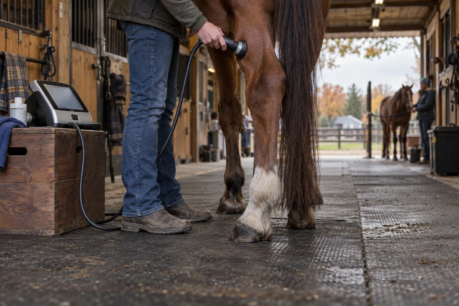
The trial followed eight adult Thoroughbred horseball horses, aged 9 to 15, each with a confirmed diagnosis of overriding dorsal spinous processes and pain in the longissimus dorsi. For practitioners working at the intersection of manual therapy and physical rehabilitation, the data points toward an adjunct modality that targets the deep stabilising layer — the very tissue group that load-dependent back care depends on.
Protocol and measurement design
Each horse received four radiofrequency treatments at two-week intervals, applied bilaterally between T9 and L6, with each session running 40 minutes. Ultrasonography captured multifidus cross-sectional area at seven sites along the thoracolumbar spine. Wearable inertial sensors recorded segmental range of motion while the horses trotted in-hand. Measurements occurred at four time points: pre-treatment, immediately after the first session, 24 hours post-session, and at eight weeks — 15 days after the fourth and final treatment.
The design deserves attention. Three post-first-treatment assessments separate acute tissue effects from delayed muscular adaptation. That timing is what permits the authors to argue for structural change rather than transient oedema or short-term fluid shifts in the muscle.
Where the muscle changed
By week eight, multifidus cross-sectional area had increased at every measured site. The largest gains appeared at T18 (+2.52 cm²), followed by L5 (+1.90 cm²) and T16 (+1.77 cm²). Progression was gradual, accumulating well after the final session — a temporal pattern consistent with muscular adaptation rather than a post-treatment swelling response.
Range of motion told a more layered story. Some increase in spinal movement appeared shortly after treatment. At eight weeks, segments of the thoracolumbar spine showed reduced movement during trot. The authors interpret this reduction as a marker of improved stabilisation — less excess motion in segments now supported by a larger multifidus.
What to verify before adding RF to a rehabilitation plan
The dataset is preliminary. Eight horses, no untreated control group, a single breed and use-case cohort. No adverse effects were reported and all animals completed the protocol, but the numbers cannot yet support claims about return-to-work timelines, ridden load tolerance, or direct comparison with manual therapy, targeted exercise, or any hands-on modality already in clinical use.
Before integrating radiofrequency into a programme, we suggest confirming three things on a case-by-case basis: a veterinary diagnosis of the underlying pathology (overriding dorsal spinous processes, with longissimus involvement in this cohort), a baseline ultrasonographic measurement of multifidus cross-sectional area, and pre-defined recovery metrics. Useful markers include change in CSA at standardised sites, change in segmental ROM under a fixed trot condition, and a defined return-to-load threshold tied to gait quality rather than calendar time. Without those benchmarks, a new modality enters the plan with no objective way to evaluate its contribution.